We are interested in the role that chromatin structure plays in gene regulation, considering both effects from packaging large domains and local effects of the nucleosome array. We work with Drosophila, combining biochemical, genetic and cytological approaches. In our initial studies, we devoted considerable effort to developing new techniques for this purpose. One such technique enables immunofluorescent staining of the polytene chromosomes to generate a distribution
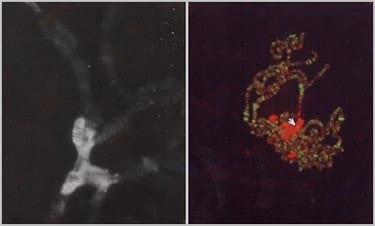
Immunofluorescent staining of the polytene chromosomes of Drosophila larvae show heterochromatin protein 1 (HP1) to be concentrated at the chromocenter and in a banded pattern along the fourth chromosome (left hand picture). Minor sites are seen along the other chromosome arms and at the telomeres. In contrast such staining shows GAGA factor, present at sites in the euchromatic arms, absent from the heterochromatic chromocenter (right hand picture). Two major sites of GAGA factor localization are seen on the fourth chromosome (arrow).
pattern for individual non-histone chromosomal proteins (Silver & Elgin, 1976). Ten years later, this technique enabled us to identify Heterochromatin Protein 1 (HP1a), a key protein for heterochromatin formation with associated gene silencing (James & Elgin, 1986; Eissenberg et al. 1990), and supported our work showing that a histone variant, H2AvD, is essential for Drosophila (van Daal & Elgin, 1986). A second approach was to use nucleases to digest the nucleosome array, followed by a Southern blot to detect the pattern for a specific gene. This technique led to the discovery of DNase I hypersensitive sites (DH sites), accessible sites within the nucleosome array (Wu et al, 1979a, 1979b), and the subsequent demonstration that such sites map to the 5’ regulatory regions of genes (C. Wu, 1980 Nature 286: 854-60; Keene et al. 1981). Further exploration found that while MNase cuts preferentially between nucleosomes, the patterns generated are often mimicked by cleavage patterns of the DNA per se (Keene & Elgin, 1981). This observation led to mapping studies using chemical as well as enzymatic reagents (e.g. Cartwright et al 1983), and ultimately sequence-level mapping of the architecture of the hsp26 gene (Thomas & Elgin, 1988).
Extensive studies of hsp26 as a test gene showed that correct assembly of the hsp26 regulatory region in an activatable form, a DH site, requires two (CT)n sites, which bind GAGA factor, as well as an immediately adjacent TFIID binding site (Gilmour et al 1989; Lu et al 1989; collaboration with the J Lis lab, Cornell University). The well-characterized hsp26 gene then proved very useful as a reporter of local chromatin structure. We have used a transposable P-element containing a copy of an hsp70-driven white gene, a visible marker for gene silencing, and a copy of hsp26 to examine the effect of insertion into different chromosomal domains. While these genes are fully active in euchromatic domains, we found that partial silencing indicative of Position Effect Variegation can be observed when the P-element is inserted into pericentric heterochromatin, telomeres, or the small fourth chromosome (Wallrath & Elgin, 1995). Investigations to examine the mechanism(s) of gene silencing have shown that changes in the local nucleosome array, as well as spatial organization in the nucleus, are both critical (e.g. Cryderman et al, 1999). Subsequent mapping of the fourth chromosome (or F element) using this reporter found that indeed most of the 1.3 Mb arm, containing ~80 genes, is packaged as heterochromatin, as inferred from the cytological analysis (Sun et al 2000, 2004). The mapping suggested that transposable element remnant 1360 is a particularly good target for heterochromatin formation; however, we found that it could only induce silencing when close to a mass of heterochromatin (Haynes et al 2006). Subsequent studies showed that 1360, while a good target, is not the only target on the fourth chromosome (Riddle et al 2008).
As noted above, earlier work in the lab identified Heterochromatin Protein 1 (HP1a) as a protein preferentially associated with the pericentric heterochromatin, and in a banded pattern along the small fourth chromosome (James & Elgin, 1986). Subsequent analysis showed that HP1 is encoded by Su(var)2-5; both mutations that would be expected to reduce the level of HP1, and a point mutation shown by others to disrupt the interaction of HP1 with H3-K9me2/3, result in suppression of Position Effect Variegation, demonstrating that HP1 plays a key role in establishing the heterochromatic structure responsible for gene silencing (Eissenberg et al 1990). HP1a proved to be highly conserved, from the yeast S. pombe to humans, and appears to play a similar role in heterochromatin formation in most metazoans (e.g. Saunders et al 1993; see Grewal & Elgin, 2002; Richards & Elgin, 2002; and Elgin & Reuter, 2013, for review). As our understanding of heterochromatin as developed, it appears that HP1 functions by stabilizing nucleosome structure (see Eissenberg & Elgin, 2014 for review).
As RNAi was reported to play a role in heterochromatin formation in fungi and plants, we wondered whether the DNA transposons such as 1360 are targeted for silencing by this system in Drosophila. In a collaborative study with the Birchler laboratory (University of Missouri), we observed that mutations in piwi, aubergine, and homeless can result in a loss of silencing at some of our reporter loci described above, as well as at other repeat loci (Pal-Bhadra et al 2004; see also Haynes et al 2006). A collaborative effort with Haifan Lin and colleagues (Yale University) provided evidence for a specific interaction between HP1a and PIWI in Drosophila melanogaster in vitro (Brower-Toland et al 2007; see also Mendez et al 2011). Further studies showed a critical role for PIWI in the female germline in assembling heterochromatin at some, but not all, TEs, although a direct interaction between HP1 and PIWI could not be verified in vivo (Wang & Elgin 2011). We then generated a P-element insertion line, #1198, in which silencing of the hsp70-white reporter requires the presence of a copy of 1360 (Sentmanat & Elgin, 2012). Genetic experiments showed that this silencing requires piwi, aub, Su(var)205 (HP1), and Su(var)3-9 (H3K9 HMT); it is also sensitive to the loss of piRNA sequences from the 1360 element, all of which argues that the piRNA system is required for targeting. In flies, the piRNA system is most active in the germ line and early embryo, and indeed a direct test showed that PIWI is required in the early embryo (demonstrated by a requirement for maternal loading), but not during larval development (demonstrated by RNAi KD), to see PEV silencing of reporter loci in the adult eye. In contrast, HP1 is required both in early embryogenesis when heterochromatin is established, and throughout development (Gu & Elgin, 2013). It appears likely that a piRNA mechanism is utilized to recognize and transposable elements and their remnants for silencing through heterochromatin formation; the pathway to HP1-H3K9me2/3 assembly may be complex,
One of the interesting features of the Drosophila genome is the extent to which the fourth (or “dot”) chromosome is packaged as heterochromatin, including that portion that codes for ~80 genes (Riddle et al 2011, 2012; Kharchenko et al 2011). This packaging has had a significant impact, resulting in genes that have a lower codon bias (presumably because of the low rate of recombination) and are significantly larger than the average, reflecting the abundance of TEs (Leung et al 2015, 2017). We are currently investigating what features of gene organization, in particular what regulatory motifs, enable fourth chromosome genes to be expressed in a chromatin domain that silences most euchromatic genes. Both a DNA manipulation strategy (swapping fragments of the hsp70-white reporter gene with fragments of a fourth chromosome gene, Rad23) and a comparative genomics approach (using phylogenetic footprinting) are being utilized, the latter work in collaboration with the Genomics Education Partnership (J Cantrell, E Gracheva, W Leung, B French, C Shaffer & SCRE, work in progress). See Riddle & Elgin, 2018, for a recent review on our understanding of the organization, evolution, and functioning of the Drosophila dot chromosome.
One of the most intriguing problems in the study of heterochromatin formation is how the cell recognizes repetitious DNA and targets it for silencing. While TE sequences are recognized through an RNAi mechanism, this is probably not the primary signal for tandem arrays, be they simple sequences or complete genes. The 1198 line discussed above has a “landing pad” site which enables us to put in (and pop out) various repetitious sequences; the insertion site is in the nesd gene (active during early embryogenesis), but close to a block of heterochromatin at the base of chromosome arm 2L. Reporter genes here are silenced if and only if a repeat – either 1360 or a tandem array – is inserted adjacent to them. We are investigating the silencing induced by GAA310 (taken from a Friedriech’s Ataxia patient, collaboration with Richard Festenstein) and by lacO256 . While the heterochromatin structure induced by GAA310 appears similar to that at pericentric heterochromatin, that induced by repeats of a bacterial sequence (lacO) has distinctive properties (E Gracheva, M Grupe, G Huang, S Bieser, & SCRE, work in progress).
Selected Publications:
Elgin, S.C.R., Boyd, J.B., Hood, L.E., Wray, W., and Wu, F.C. (1974) “A prologue to the study of the nonhistone chromosomal proteins,” Cold Spring Harbor Symp. Quant. Biol. 38, 821-833.
Elgin, S.C.R., and Weintraub, H. (1975) “Chromosomal proteins and chromatin structure,” Ann. Rev. Biochem. 44, 725-74.
Silver, L.M., and Elgin, S.C.R. (1976) “A method for determination of the in situ distribution of chromosomal proteins,” Proc. Nat. Acad. Sci. USA 73, 423-27.
Elgin, S.C.R., Serunian, L.A., and Silver, L.M. (1978) “Distribution patterns of Drosophila nonhistone chromosomal proteins,” Cold Spring Harbor Symp. Quant. Biol. 42, 839-50.
Wu, C., Bingham, P.M., Livak, K.J., Holmgren, R. and Elgin, S.C.R. (1979a) “The chromatin structure of specific genes. I. Evidence for higher order domains of defined DNA sequence,” Cell 16, 797-806.
Wu, C., Wong, Y.C. and Elgin, S.C.R. (1979b) “The chromatin structure of specific genes. II. Disruption of chromatin structure during gene activity,” Cell 16, 807-14.
Keene, M.A., Corces, V., Lowenhaupt, K., and Elgin, S.C.R. (1981) “DNase I hypersensitive sites in Drosophila chromatin occur at the 5′ ends of transcribed regions,” Proc. Natl. Acad. Sci. USA. 78, 143-46.
Keene, M.A., and Elgin, S.C.R. (1981) “Micrococcal nuclease as a probe of DNA sequence organization and chromatin structure,” Cell 27, 57-64.
Elgin, S.C.R. (1982) “Chromatin Structure, DNA Structure,” (News and Views), Nature 300, 402-03.
Elgin, S.C.R., Cartwright, J.L., Fleischmann, G., Lowenhaupt, K., and Keene, M.A. (1983) “Cleavage reagents as probes of DNA sequence organization and chromatin structure: Drosophila melanogaster Locus 67B1,” Cold Spring Harbor Symp. Quant. Biol. 47, 529-538.
Cartwright, I.L., Hertzberg, R.P., Devan, P.B., and Elgin, S.C.R. (1983) “Recognition of the nucleosomal structure of chromatin by a cleavage reagent with low sequence preference: (Methidiumpropyl-EDTA) iron (II),” Proc. Natl. Acad. Sci. USA, 80, 3212-3217.
Cartwright, I.L. and Elgin, S.C.R. (1984) “Chemical footprinting of 5S RNA chromatin in embryos of Drosophila melanogaster,” EMBO J. 3, 3101-3108.
Flick, J.T., Eissenberg, J.C., and Elgin, S.C.R. (1986) “Micrococcal nuclease as a DNA structural probe: Its recognition sequences, their genomic distribution, and correlation with DNA structure determinants,” J. Mol. Biol. 190, 619-633.
James, T.C. and Elgin, S.C.R. (1986) “Identification of a nonhistone chromosomal protein associated with heterochromatin in Drosophila melanogaster and its gene,” Molec. Cell. Biol., 6, 3862-3872.
Gilmour, D.S. and Elgin, S.C.R. (1987) “Localization of specific topoisomerase I interactions within the transcribed region of active heat shock genes using the inhibitor camptothecin,” Molec. Cell Biol. 7, 141-148.
Thomas, G.H. and Elgin, S.C.R. (1988) “Protein/DNA architecture of the DNase I hypersensitive region of the Drosophila hsp26 promoter,” EMBO J., 7, 2191-2201.
Elgin, S.C.R. (1988) “The formation and function of DNase I hypersensitive sites in the process of gene activation,” J. Biol. Chem. 263, 1259-62.
Gilmour, D.S., Thomas, G.H., and Elgin, S.C.R. (1989) “Drosophila nuclear proteins bind to regions of alternating C and T residues in gene promoters,” Science 245, 1487-1490.
Eissenberg, J.C., James, T.C., Foster-Hartnett, D.M., Hartnett, T., Ngan, V., and Elgin, S.C.R. (1990) “A mutation in a heterochromatin-specific chromosomal protein is associated with suppression of position effect variegation in Drosophila melanogaster,” Proc. Natl. Acad. Sci. USA 87, 9923-9927.
van Daal, A. and Elgin, S.C.R. (1992) “A histone variant, H2AvD, is essential in Drosophila melanogaster,” Molec. Biol. Cell. 3, 593-602.
Lu, Qin, Wallrath, L.L, Allen, B.D., Glaser, R.L., Lis, J.T., and Elgin, S.C.R. (1992) “A promoter sequence containing (CT)n•(GA)n repeats is critical for the formation of the DNase 1 hypersensitive sites in the Drosophila hsp26 gene,” J. Mol. Biol. 225, 985-998.
Saunders, W.S., Chue, C., Goebl, M., Craig, C., Clark, R.F., Powers, J.A., Eissenberg, J.C., Elgin, S.C.R., Rothfield, N.F., and Earnshaw, W.C. (1993) “Molecular cloning of a human homologue of Drosophila heterochromatin protein HP1 using anticentromere autoantibodies with anti-chromo specificity,” J. Cell Sci. 104, 573-582.
Elgin, S.C.R., Granok, H., Lu, Q., and Wallrath, L.L. (1994) “The role of chromatin structure in regulating gene expression: The hsp26 gene of Drosophila melanogaster,” Cold Spring Harbor Symp. Quant. Biol. 58, 83-96.
Elgin, S.C.R., editor (1995) Chromatin Structure and Gene Expression, Oxford University Press, New York.
Wallrath, L.L., and Elgin, S.C.R. (1995) “Position effect variegation in Drosophila is associated with an altered chromatin structure,” Genes & Develop. 9, 1263-1277.
Elgin, S.C.R. (1996) “Heterochromatin and gene regulation in Drosophila,” Curr. Opn. Genet. Develop. 6, 193-202.
Cryderman, D.E., Morris, E.J., Biessman, H., Elgin, S.C.R., and Wallrath, L.L. (1999) “Silencing at Drosophila telomeres: nuclear organization and chromatin structure play critical roles,” EMBO J. 18, 3724-3735.
Sun, F.-L., Cuaycong, M.H., Craig, C.A., Wallrath, L.L., Locke, J., and Elgin, S.C.R. (2000) “The fourth chromosome of Drosophila melanogaster: interspersed euchromatic and heterochromatic domains,” Proc. Natl. Acad. Sci. USA 97, 5340-5345.
Elgin, S.C.R. and Workman, J. J., editors (2000) Chromatin Structure and Gene Expression, second edition, Oxford University Press, New York.
Sun, F.-L., Cuaycong, M.H., and Elgin, S.C.R. (2001) “Long-range nucleosome ordering is associated with gene silencing in Drosophila melanogaster pericentric heterochromatin,” Mol. Cell. Biol. 21, 2867-2879.
Grewal, S.I.S., and Elgin, S.C.R. (2002) “Heterochromatin: New Possibilities for the Inheritance of Structure,” Curr. Opin. Genet. Develop. 12, 178-187.
Richards, E.J., and Elgin, S.C.R. (2002) “Epigenetic Codes for Heterochromatin Formation and Silencing: Rounding up the Usual Suspects,” Cell 108, 489-500.
Shaffer, C.D., Stephens, G.E., Thompson, B.A., Funches, L., Bernat, J.A., Craig, C.A., and Elgin, S.C.R. (2002) “Heterochromatin protein 2 (HP2), a partner of HP1 in Drosophila heterochromatin,” Proc. Natl. Acad. Sci. USA 99, 14322-14337.
Lu, Q., Teare, J.M., Granok, H., Swede, M.J., Xu, J. and Elgin, S.C.R. (2003) “The capacity to form H-DNA cannot substitute for GAGA factor binding to a (CT)n•(GA)n regulatory site,” Nucleic Acids Res. 31, 2483-2494.
Pal-Bhadra, M., B. A. Leibovitch, S. G. Gandhi, M. Rao, U. Bhadra, J. A. Birchler, S. C. R. Elgin (2004) “Heterochromatic silencing and HP1 localization in Drosophila are dependent on the RNAi machinery,” Science 303, 669-672.
Sun, F.-L., Haynes, K., Simpson, C.L., Lee, S.D., Collins, L., Wuller, J., Eissenberg, J.C., and Elgin, S.C.R. (2004) “Cis-acting determinants of heterochromatic formation on Drosophila melanogaster chromosome four,” Mol. Cell. Biol., 24, 8210-8220.
Haynes, K.A., Leibovitch, B.A., Rangwala, S.H., Craig, C., and Elgin, S.C.R. (2004) “Analyzing heterochromatin formation using chromosome four of Drosophila melanogaster,” Cold Spring Harbor Symp. Quant. Biol. 69, 267-272.
Huisinga, K.L., Brower-Toland, B., and Elgin, S.C.R. (2006) “The contradictory definitions of heterochromatin: transcription and silencing.” Chromosoma 115, 110-122.
Haynes, K.A., Caudy, A.A., Collins, L. and Elgin S.C.R. (2006) “Element 1360 and components of the RNAi system contribute to HP1-dependent silencing of a pericentric reporter,” Current Biology 16: 2222-27.
Brower-Toland, B., Findley, S.D., Jiang, L., Liu, L., Yin, H., Dus, M., Zhou, P., Elgin, S.C.R., and Lin, H. (2007) “Drosophila PIWI associates with chromatin and interacts directly with HP1a,” Genes & Develop. 21: 2300-2311.
Grewal, S.I.S., and Elgin, S.C.R. (2007) “Transcription and RNA interference in the formation of heterochromatin,” Nature 447: 399-406.
Riddle, N.C., Leung, W., Haynes, K.A., Granok, H., Wuller, J, and Elgin, S.C.R. (2008) “An investigation of heterochromatin domains on the fourth chromosome of Drosophila melanogaster.” Genetics 178: 1177-1191. PMCID: PMC2278077.
Huisinga, K., and Elgin, S.C.R, (2009) “Small RNA directed heterochromatin formation in the context of development: what flies might learn from fission yeast.” Biochem. Biophys. Acta 1789: 3-16. PMCID: PMC2633771.
Pope, WH, ….Elgin, SCR, …Hatfull, GF (2011) Expanding the diversity of mycobacteriophages: Insights into genome architecture and evolution. PLoS One 6: e16329. http://dx.plos.org/10.1371/journal.pone.0016329 PMCID: PMC3029335.
Riddle, NC, A Minoda, PV Kharchenko, AA Alekseyenko, YB Schwartz, MY Tolstorukov, AA Gorchakov, C Kennedy, D Linder-Basso, JD Jaffe, G Shanower, MI Kuroda, V Pirrotta, PJ Park, SCR Elgin, GH Karpen (2011) “Plasticity in patterns of histone modifications and chromosomal proteins in the Drosophila heterochromatin,” Genome Res. 21:147-63. PMID: 21177972.
Kharchenko, PV, AA Alekseyenko, YB Schwartz, A Minoda, NC.Riddle, J Ernst, PJ Sabo, E Larschan, AA Gorchakov, T Gu, D Linder-Basso, A Plachetka, G Shanower, MY Tolstorukov, LJ Luquette, R Xi, YL Jung, R Park, EP Bishop, TP Canfield, R Sandstrom, RE Thurman, DM MacAlpine, J Stamatoyannopoulos, M Kellis, SCR Elgin, MI Kuroda, V Pirrotta, G Karpen, PJ Park. (2011) Comprehensive analysis of the chromatin landscape in Drosophila melanogaster. Nature 471: 480-5. PMID: 21179089. PMCID: PMC3109908.
Mendez, DL, D Kim, M Chruszcz, GE Stephens, W Minor, S Khorasanizadeh, and SCR Elgin (2011) The HP1a disordered C-terminus and Chromo Shadow Domain cooperate to select target peptide partners. ChemBioChem 12: 1084-96. PMCID: PMC3154745.
Wang, S H, and Elgin, SCR (2011) “Drosophila Piwi functions downstream of piRNA production mediating a chromatin-based silencing mechanism in female germline,” Proc Natl Acad Sci USA. 108: 21164-69. PMCID: PMC3248523.
Riddle, NC, YL Jung, T Gu, AA Alekseyenko, D Asker, H Gui, PV Kharchenko, A Minoda, A Plachetka, YB Schwartz, MY Tolstorukov, MI Kuroda, V Pirrotta, GH Karpen, PJ Park, SCR Elgin. (2012) “Enrichment of HP1a on Drosophila chromosome 4 genes creates an alternative chromatin structure critical for regulation in this heterochromatic domain,” PLoS Genetics 8: e1002954 PMCID: PMC3447959.
Sentmanat, M, and SCR Elgin. (2012) Ectopic assembly of heterochromatin in Drosophila triggered by transposable elements. Proc Natl Acad Sci USA 109: 14104-9. PMCID: PMC3435190.
Elgin, S.C.R. and Reuter, G. (2013) “Position-effect variegation, heterochromatin formation, and gene silencing in Drosophila,” in “Epigenetics,” 2nd edition, ed. C.D. Allis, T. Jenuwein, & D. Reinberg, Cold Spring Harbor Laboratory Press, NY (2015). Published in Cold Spring Harbor Perspect Biol Aug 1:5(8). PMID: 23906716.
Gu, T, and Elgin, SCR (2013) Maternal delpletion of Piwi, a component of the RNAi system, impacts heterochromatin formation in Drosophila. PLoS Genetics 9: e1003780/ PMCID: PMC3777992.
Ho, JW, …Gu T, …, Riddle NC, …Elgin SC, Park PJ (2014) Comparative analysis of metazoan chromatin organization. Nature 512: 449-52. PMID: 25164756.
Eissenberg, JC & SCR Elgin (2014) HP1a: A structural chromosomal protein regulating transcription. Tr Genetics 30: 103-10. PMCID: PMC3991861.
Leung, W….[940 students, 72 faculty]…Elgin, SCR (2015) The Drosophila Muller F elements maintain a distinct set of genomics properties over 40 million years of evolution. G3: GENES, GENOMES, GENETICS 5: 719-740. PMCID: PIMC4426361.
NOTE: this paper received considerable social commentary because of the high number of undergraduate co-authors; see
http://gep.wustl.edu/community/four_genomes_paper for a summary.
Huisinga KL, NC Riddle, W Leung, S Shimonovich, S McDaniel, A Figueroa-Clarevega, SCR Elgin (2016) Targeting of P element reporters to heterochromatic domains by transposable element 1360 in Drosophila melanogaster. Genetics 202: 565-82. PMID: 26680659.
Leung, W…SCR Elgin. (2017) Retrotransposons are the major contributors to the expansion of the Drosophila ananassae Muller F element. G3 7: 2439-60. doi: 10.1534/g3.117.040907. PMID:28667019. (This paper from the Genomics Education Partnership has 276 co-authors; 239 of them contributed as students.)
Leung, W, & SCR Elgin (2018) Response to the Letter to the Editor by Hotopp and Klasson. G3 8: 375. Doi: https://doi.org/10.1534/g3.117.300379.
Riddle, NC and SCR Elgin (2018) The Drosophila dot chromosome: where genes flourish amidst repeats. Genetics 210: 757-772. doi: 10.1534/genetics.118.301146 PMID: 30401762 (FlyBook chapter).
Liu, Y, L Sargent, W Leung, SCR Elgin, J Goecks (2018) G-OnRamp: A Galaxy-based platform for creating genome browsers for collaborative genome annotation. In preparation.