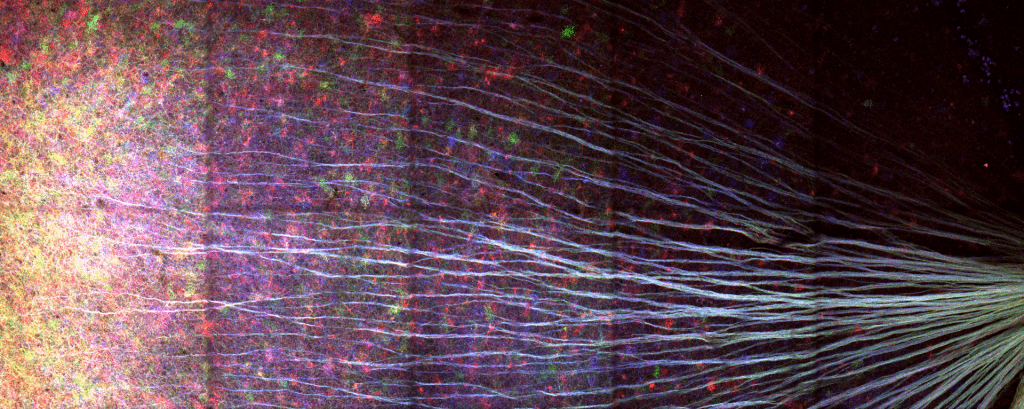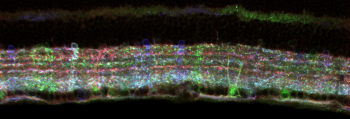

AAV-Brainbow labeling of retinal neurons in wholemount (top) and cross sections (right) showing the diversity of retinal neurons in the inner retina.


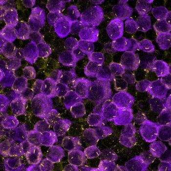
Retinal ganglion cell IGF1 receptor localization to primary cilia is critical for IGF1 induced regeneration. Our lab is studying the role of primary cilia in both RGC axon regeneration and how primary cilia regulate neuromodulator signaling.

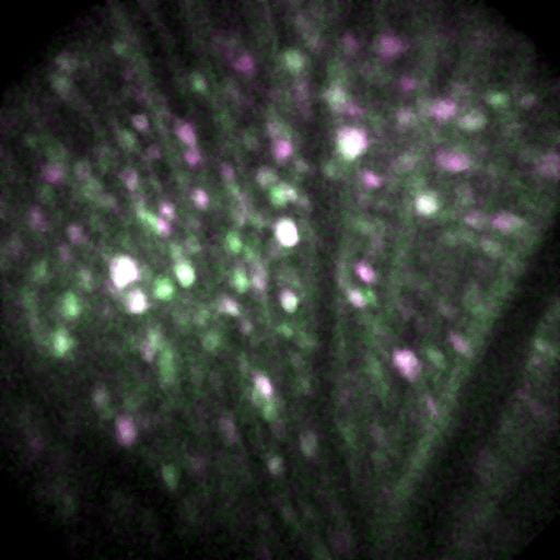
In vivo biosensor imaging of retinal ganglion cells demonstrates different fundamental properties across this family of neurons. We are investigating how these differences impact survival during neurodegeneration.

Our lab celebrates events with homemade ice cream. We’ve made over 50 different flavors. Some of the best include Pretzel, Maple Bacon, Peanut Butter Jelly, Mango & Chorizo, Pad Thai, Rice, Grapefruit & Caramelized White Chocolate, and Earl Grey Tea with Sandwich Cookies.