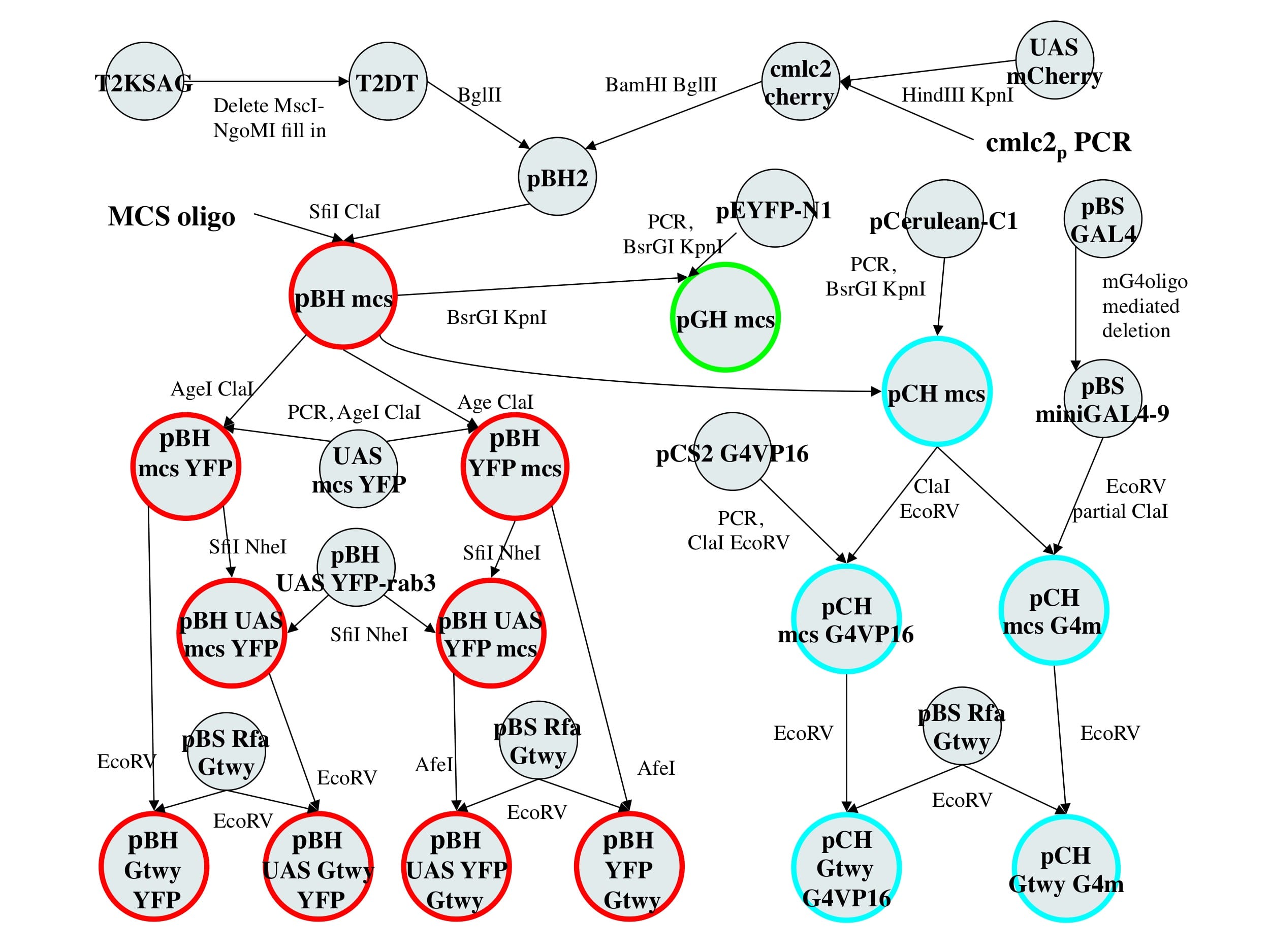
| pBleeding Heart construction The vectors are based on the tol2 vector T2KSAG (ref 1). We designed the vectors to maintain unique sites at as many junctions as possible to allow for simple swapping of componenets. Vector construction was done as follows (flow diagram):
|
| pCold Heart construction
|
pGep Heart construction
|
| References 1) Kawakami K, Takeda H, Kawakami N, Kobayashi M, Matsuda N, Mishina M. A transposon-mediated gene trap approach identifies developmentally regulated genes in zebrafish. Dev Cell. 2004 Jul;7(1):133-44. 2) Huang CJ, Tu CT, Hsiao CD, Hsieh FJ, Tsai HJ.Germ-line transmission of a myocardium-specific GFP transgene reveals critical regulatory elements in the cardiac myosin light chain 2 promoter of zebrafish. Dev Dyn. 2003 Sep;228(1):30-40. 3)Shaner NC, Campbell RE, Steinbach PA, Giepmans BN, Palmer AE, Tsien RY. Improved monomeric red, orange and yellow fluorescent proteins derived from Discosoma sp. red fluorescent protein. NAt Biotechnol. 2004 Dec;22(12):1567-72. 4) from Clontech, Inc. 5) The sequenced UAS contains 11 copies of the GAL4 binding domain. The UAS was derived from pCS2-GAL4VP16 obtained from R. Wong Lab (WUSTL) which originally contained 14 sites. This UAS repeat appears to be somewhat unstable. 6) Walhout AJ, Temple GF, Brasch MA, Hartley JL, Lorson MA, van den Heuvel S, Vidal M. GATEWAY recombinational cloning: application to the cloning of large numbers of open reading frames or ORFeomes. Methods Enzymol. 2000;328:575-92. 7) Rizzo MA, Springer GH, Granada B, Piston DW. An improved cyan fluorescent protein variant useful for FRET. Nat Biotechnol. 2004 Apr;22(4):445-9. 8) Sadowski I, Ma J, Triezenberg S, Ptashne M. GAL4-VP16 is an unusually potent transcriptional activator. Nature. 1988 Oct 6;335(6190):563-4. 9) Ding WV, Johnston SA. The DNA binding and activation domains of Gal4p are sufficient for conveying its regulatory signals.Mol Cell Biol. 1997 May;17(5):2538-49. |