Our Experimental Approach
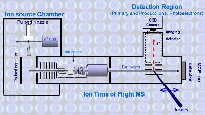 Anions are produced in the source chamber, which is a cylindrical stainless steel chamber held at approximately 10-5 torr during operation by an 8000 L/s diffusion pump (Leybold DIP8000, backed by a Leybold DB40 mechancical pump). A high pressure of gas mixture containing inert carrier (usually Ar) seeded with precursor molecules is expanded into the source chamber through a pulsed nozzle (General Valve Series 9), typically at a repetition rate of 10 Hz. The adiabatic expansion creates the conditions for cluster formation. The expansion either passes through a pulsed discharge or is crossed by a 1 keV electron beam (Kimball Physics EGPS 1017) and a mixture of anions, cations and neutral species are produced. The anions are extracted into a Wiley McLaren time of flight mass spectrometer using the pulsed repeller, which is negatively charged. The volatage can be varied to set the optimum Wiley McLaren focusing conditions and the time of application of the pulse to the repeller sets the zero for the ion time of flight. Fig. 1 Schematic of Anion Imaging Spectrometer
As the ions enter the time of flight tube they are accelerated through approx 2 kV and the anions can be steered and focussed by means of electrostatic deflectors and an einzel lens. The ions enter a cylindrical stainless steel tube, the potential of which is rapidly switched from 2000 to 0 V (a potential switch) to rereference to ions to ground and alleviate the need to float the whole instrument at 2 kV. The time of flight region is separated from the source chamber and detection regions by small apertures and is maintained at 10-8 torr by two 360 L/s turbo pumps (Leybold TMP 361, backed by a Leybold DB16 rotary pump). The detection region can be further isolated from the central section of the instrument by a 6 inch gate valve which is kept closed when not in operation. This and the action of a further turbo pump maintains the pressure in the detection region at <5×10⁻⁹ torr.
At the end of the flight tube ions are detected using a microchannel plate (MCP) detector and the mass sepctrum is recorded using a four channel digital oscilloscope (LeCroy Wavejet WJ334 350). Within the detection region the anions pass between the bottom two electrodes of a three electrode velocity mapped imaging arrangement. A laser pulse is synchronized to coincide with the ion mass of interest using a digital delay generator (Berkley Nucleonics Corp. BNC 505). Either ns (Spectra Physics Syrah pumped by an INDI10 Nd:YAG laser) or fs (Spectra Physics Spitfire Pro regeneratively amplifying a Spectra Physics Tsunami laser) laser pulses are available. The ns system allows tuning throughout the visible range of the spectrum and into the ultra violet via means of frequency doubling. The fs system also allows tuning around the fundamental of the Ti:Sapphire medium and we have the capacity to frequency double, triple and quadruple. Further tuning is also possible using a Spectra Physics OPA.
Electrons detached from the anions within the imaging arrangement are forced onto a position sensitive detector which lies at the end of a 20 cm, mu metal shielded, field free electron flight tube. The detector (Burle Inc.) consists of a pair of matched imaging quality MCPs backed by a (P20) phosphor screen. The bias across the MCPs is pulsed high (approx 2 kV) for a 200 ns window around the arrival of the laser pulse to discriminate agains background noise. Electron impacts on the MCP, which cause bright flashes on the phosphor screen, are recorded using an IMPERX 240 CCD camera and accumulated frame by frame.
Controls and Data Collection

Optical Bench

Ion TOF and Detection Region

Source Chamber and TOF

Ultra-fast System

Ion Source Chamber
Dye Laser

Controls
